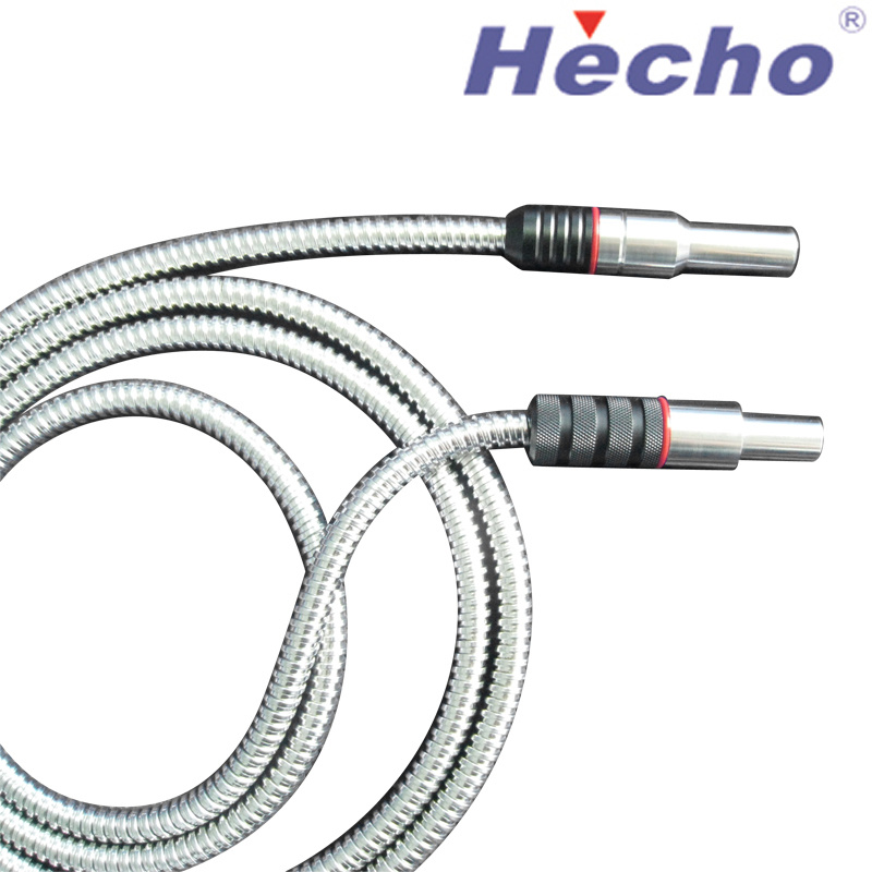
Insertion Tube Endoscope
The insertion tube endoscope is the section of an endoscope that is inserted into the body cavity for examination. It is used to inspect the stomach, duodenum, small intestine or large intestine.
The insertion tube of an endoscope is made of a flexible elongated structural body, with an outer cover on the outer periphery. The outer cover serves to prevent body fluids from entering the inside of the insertion tube.
The Light Bundle
An insertion tube endoscope is a type of medical instrument that is used to visualize and diagnose the interior of the human body. They can be used for a variety of medical procedures, such as diagnosing and removing tumours from the lungs and digestive tract, taking samples of tissue (biopsy), placing tubes (stents) through blockages in the bile duct, oesophagus, duodenum or colon, etc.
Most of the time, the insertion tube endoscope is inserted through one of the body’s natural openings, such as the mouth, urethra, or anus. The endoscope contains a light source, air pump and video processor. The video camera is positioned at the distal tip of the insertion tube and magnifies what is inside the body.
The insertion tube endoscope is also equipped with a deflectable portion on the distal tip, which can be manipulated by an endoscopist. This portion is referred to as the bending section and is constructed differently from the rest of the insertion tube.
In a typical endoscope, the bending section is composed of a series of oddly-shaped metal rings, each ring connected to the next ring on the bending section. These rings are placed around the outside of the insertion tube and can be rotated by an endoscopist using the control knob to bend the bending section and the distal tip.
When the bending section is deflected, the insertion tube ends up angled in line with the angulation dials on the scope body. To maintain the angulation, steel wires are used to move the end of the insertion tube in line with these angulation dials. These wires can stretch over time and should be operated carefully.
As a result, it is difficult to ensure that the proximal end of an optical fiber bundle is not bent when it is passed through the bending portion. Therefore, it is necessary to develop a suitable assembling method for the insertion section of the endoscope that can ensure the proximal end of an optical fibre bundle is not bent when the insertion tube is curved during the use of the endoscope.
The Image Bundle
The Image Bundle is a continuous strand of flexible glass fibers that transmits the image through the scope to the eyepiece. It also carries light to illuminate the area being examined by the insertion tube endoscope. A video endoscope is a bit different from a fiberscope in that the image bundle is replaced by a lens system that reduces and focuses the image to the surface of a charge-coupled device (CCD) chip.
To produce a fiberoptic image, the individual glass fibers in an imaging bundle must be arranged in a coherent order, which is achieved by fusing the individual fiber faces together at each end of the bundle. This is very difficult and time consuming to do, as each fiber needs to be carefully aligned in order to achieve the desired resolution.
Despite the difficulty, fiberoptic imaging is often used for observation of small, complex structures in medical and veterinary settings. This type of imaging is used for a wide variety of applications, including diagnosis and insertion tube endoscope treatment of diseases such as cancer, fibrosis, and infectious diseases, as well as for assisting surgeons in performing a variety of procedures.
A typical fiberscope has a control section and an insertion section, which extends from the control section to the distal end member. The optical fiber bundles are passed between the control section and the insertion section. The fiber bundles include an image guide, a light guide, operating wires, and forceps channel.
In the insertion tube, the optical fiber bundles are passed through a series of bores in a flexible tube that is in turn inserted into the body cavity or other similar location. The insertion tube is usually made of a flexible resin, such as polyurethane or polyvinyl chloride.
This flexible tube can be made in a number of sizes. It is generally a multi-lumen tube with a plurality of axial bores that are adapted to receive optical fiber bundles, operating wires, forceps channels, and other components.
One of the most vulnerable parts of a scope is the bending section, which is located in the distal tip of the insertion tube. This area is incredibly susceptible to damage when fluid invades the bending rubber that makes up this section of the tube. This is a major reason why the bending section should be protected from any external pressure or moisture.
The Universal Cord
The Universal Cord is an important component that runs from the light guide connector control boot down to the power supply, so it needs to be sturdy. It also has a strain relief boot at the upper end of the light guide cord and both ends of the insertion tube, which helps prevent the connection from “buckling” under heavy loads.
The Light guide connector (LGC) is an area of the scope that plugs into the processor to allow light to enter the scope during a procedure. The LGC is comprised of quite a few components, but most of them are very robust and resistant to physical damage.
However, one section of the light guide connector is susceptible to damage: the suction channel that runs from the suction tree on the LGC down to the control handle. When this suction channel gets kinked, it can cause the scope to not work properly.
This can be an issue because the suction channel is hollow, so it can buckle and kink when it’s twisted or moved during a procedure. If it’s kinked 32cm from the control boot, it can impede the aeration/perfusion tube and the suction tube that run from the treatment appliance passage on the LGC down to the insertion unit on the control handle.
If this happens, the aeration/perfusion tube can’t be hooked up properly, which could lead to poor performance during a procedure. This can also cause the suction tube to get kinked and can damage the light bundle that’s carrying the image or the fiber optic light bundle that’s carrying the light.
Since the light bundle and suction channel are both run through the Universal Cord, it’s important that the universal cord doesn’t kink or buckle so that the light guide connector and the control handle can work properly during a procedure. If the universal cord kinks or buckles, it can impede both of these critical areas, which is why it’s so important to take care of this part as soon as possible.
The universal cord is an essential part of an insertion tube endoscope because it allows the scope to be used in an angled position. It also makes the insertion tube more maneuverable and easier to insert into the patient. This can help the patient’s recovery and make for a more pleasant experience during the procedure.
The Insertion Tube
The insertion tube of an endoscope is the long, flexible tube that is inserted into the patient’s body during normal use. It contains bundles of optical fibres that carry light down the tube into the body, and a video camera on the other end that magnifies and projects the image onto a television screen for the physician to see.
The lens of the endoscope is a circular glass that is used to focus a miniature image of the GI mucosa and to project the images back to the physician’s eyepiece or into a video camera. The insertion tube also contains a small tube that carries water and air down into the endoscope.
When the endoscope is inserted into the intestine, the water/air tubes merge together into a single tube just a insertion tube endoscope few inches from the distal tip of the instrument (see Fig. 3.2). The nozzle of the combined air/water tube is positioned just a few inches from the objective lens on the endoscope’s tip. This nozzle is controlled by the endoscopist and can be used to direct water across the lens or to insufflate the patient’s GI tract with air.
In addition, the insertion tube of the endoscope often has a biopsy channel that allows passage of biopsy forceps and other tools that may be needed during therapy. The opening for this is covered by a biopsy cap that creates an air-tight seal to ensure that the channel does not leak when the instrument is moved from the insertion tube into the biopsy channel.
Another useful feature of an insertion tube is its ability to be “torqued” or flexed. In general, the insertion tube of an endoscope should have the strength to withstand repeated bending and torqueing from manipulation by the surgeon or the patient.
The shaft of the insertion tube of an endoscope can be constructed from multiple materials and with different strengths and characteristics. The goal of the design process is to combine flexibility, elasticity, column strength, and torquing capability to give the endoscopist the best possible handling experience with the insertion tube.